Part of the Oxford Instruments Group
Expand

Cypher Family AFMs

Proven AFMs engineered for exceptional resolution, speed, and versatility

The Cypher Family delivers industry-leading AFM performance built on an ultra‑stable, low‑noise platform - enabling fast,& high‑resolution imaging and broad research versatility across materials science and life science disciplines.

Request Pricing
Cypher Family AFMs

Why Cypher AFM

The Cypher AFM Family consists of four models: the Cypher L, Cypher S, Cypher ES, and Cypher VRS1250. Each model builds upon the capabilities of the preceding models, enabling you to choose the configuration that best suits your research and budget while also providing a path for future upgrades. Every model shares the same strong core foundation of performance, versatility, and ease of use.

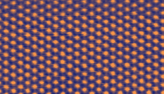
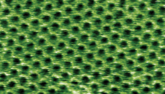
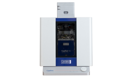
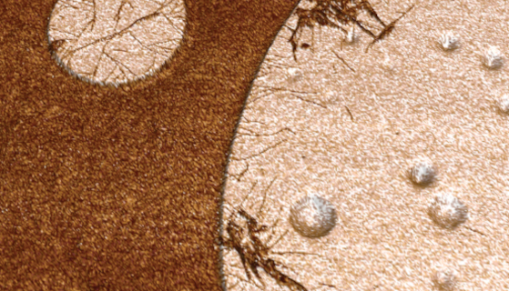
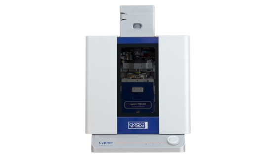
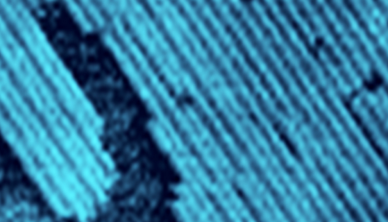
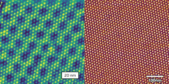
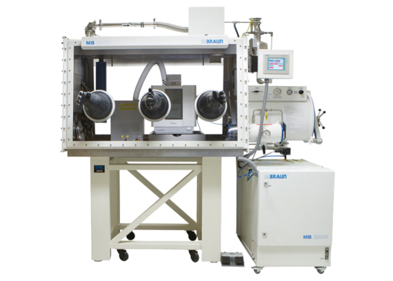
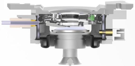
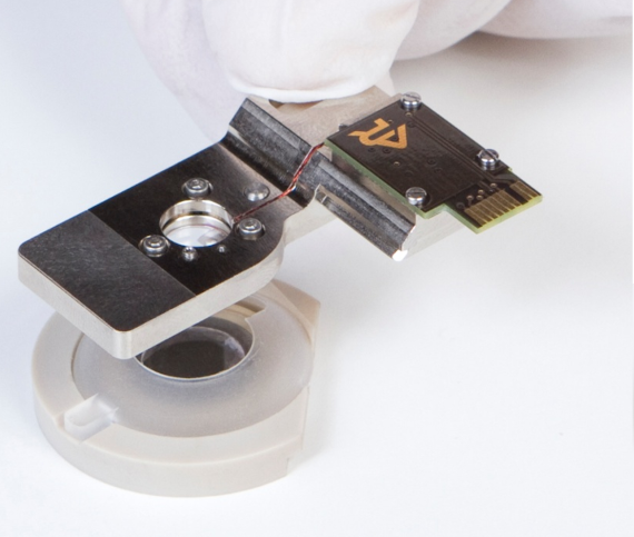
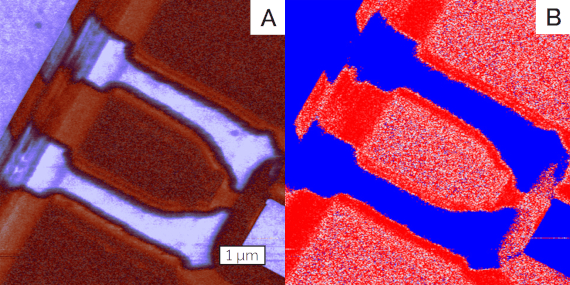
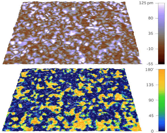
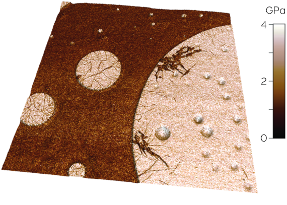
Routinely achieve higher resolution
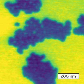
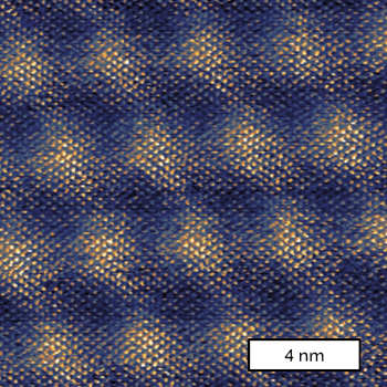
A compact ultra-stable design with a noise floor 50% lower than most competitors easily resolves molecular or atomic features that are difficult or impossible to see on typical AFMs.
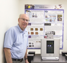
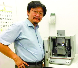
Exceptional research versatility
Enabled by the industry’s widest range of operating modes and modular accessories supporting electrical, mechanical, functional, and environmental studies across materials science and life science applications.

Fast scanning comes standard
Cypher L, S and ES acquire images 10-40× faster than conventional AFMs, while our Cypher VRS1250 delivers true video-rate imaging up to 45 frames/s.

Productivity-boosting ease of use
Motorized setup and operation, optional blueDrive photothermal excitation, and streamlined workflows in our Ergo operating software with advanced AutoPilot algorithms help every user achieve outstanding results.

Explore Cypher

The most accessible entry point to world class AFM performance, providing high-resolution core imaging today with a future-proof upgrade path to more advanced Cypher models.

The gold standard for ultra high performance AFM, offering <15 pm noise floor, up to 40× faster scanning, and comprehensive electrical, mechanical, and functional imaging modes—ideal for 2D materials and piezo/ferroelectric materials research.

The first AFM engineered specifically for exceptional environmental control, featuring a fully-sealed sample chamber with modular options for operation in controlled gases or liquids, sample heating and cooling, humidity control, and electrochemistry. Perfect for investigating polymers, energy materials, surface chemistry, biomolecules, and biomembranes.

The first and only full-featured video-rate AFM, capturing images at up to 45 frames/s while maintaining unrivaled versatility. Break new ground visualizing nanoscale dynamics and empower your group to tackle the most demanding multidisciplinary research

Hear How Cypher AFMs Make a Difference

  • Since we got the Cypher we are glued to it. We love the resolution, the reliability and… the blueDrive! It is allowing us to make quantitative measurements on a whole range of biological systems from single molecules to plant tissues. The Cypher is inspiring us to try many things that we would have not tried otherwise.

    Professor Sonia Contera, University of Oxford, United Kingdom

    Professor Sonia Contera, University of Oxford, United Kingdom
  • Cypher is truly the first third-generation AFM. The Asylum Research folks have looked at everything that limited AFM performance in the past and addressed all of them in this new design. Cypher has better resolution, less noise, less drift, is more accurate, is able to scan faster and, on top of that, is the easiest to use AFM I have ever seen.

    Professor Bruce Parkinson, University of Wyoming (USA)

    Professor Bruce Parkinson, University of Wyoming (USA)
  • Asylum’s Cypher is clearly the best choice for polymer research. When I compared Cypher to the alternatives, it became very obvious that Asylum Research is far ahead of their competitors in pushing the limits of AFM performance and developing tools for nanomechanical measurements.

    Professor Ken Nakajima, Tokyo Institute of Technology (Japan)

    Professor Ken Nakajima, Tokyo Institute of Technology (Japan)
  • Cypher is one fantastic AFM. It sets the standard for both resolution and ease of use. It makes routine characterization easy and opens many possibilities for high resolution imaging. We're really excited to have this instrument in our lab.

    Professor Ray Dagastine, University of Melbourne (Australia)

    Professor Ray Dagastine, University of Melbourne (Australia)
  • Cypher is by far the best, most stable and most configurable AFM I have ever used.

    Andras Kis, EPFL Switzerland

    Andras Kis, EPFL Switzerland

View our Product range

= Optional

Related Applications

Cypher AFMs are widely used by leading research groups for the fabrication and characterization of van der Waals heterostructures. Their exceptional stability, high resolution, and fast-scanning performance make them ideal for common use cases, including:

  • Precise nanolithography tools for cutting 2D materials during fabrication.
  • Simple, consistent visualization of moiré patterns for twist-angle determination in stacked structures.
  • Exceptional performance inside a glovebox for work with air- and water-sensitive materials

Recommendation: Cypher L or Cypher S are most commonly used.

Moiré patterns formed by twisted bilayer graphene on hexagonal boron nitrade, imaged using conductive AFM on a Cypher S. Image courtesy of S. Zhang, Tsinghua University.

Moiré patterns formed by near-magic-angle twisted bilayer graphene, imaged using Torsional Force Microscopy on a Cypher S Sample. Image courtesy of Young Lab, University California Santa Barbara.

Scientists at Oxford Instruments Asylum Research, in collaboration with leading research groups, have developed powerful AFM tools for characterizing piezo- and ferro-electric materials. Cypher AFMs, with their ultra-low-noise performance and exceptional versatility, enable:

  • High-sensitivity piezoresponse force microscopy (PFM) using patented DART PFM for reliable out-of-plane and in-plane domain imaging.
  • Comprehensive ferroelectric material analysis with Switching Spectroscopy PFM and box-in-box poling for measuring coercive fields, “wake-up” effects, and domain stability.
  • Safe, integrated high-voltage tip-bias for low-response piezoelectrics or high-switching-field ferroelectrics.

Recommendation: Cypher S (See also Vero S for even more advanced PFM capabilities).

DART PFM images of multiferroic BiFeO3 nanofibers, 1 µm scams. The amplitude image on the left shows the magnitude of the vertical response. The phase image on the right shows the polarization of the response. Collaboration with S. Xie, Xiangtan University and J.Y. Li, University of Washington.

Cypher AFMs are optimized for polymer research and R&D, combining ultra‑low noise, advanced nanomechanical modes, and precise environmental control for formulation, composite development, and process optimization. They enable:

  • High‑resolution imaging of polymer morphology, including blend domains, interphases, filler dispersion, and lamellar structure.
  • Quantitative nanomechanical mapping (AM‑FM, Fast Force Mapping, contact resonance) to measure modulus and viscoelastic properties.
  • In situ thermal and environmental studies with integrated heating/cooling and sealed liquid or gas environments to track structural changes.

Recommendation: Cypher ES Polymer Edition includes a package of commonly used tools for polymer research.

Crystalline lamella in polyethylene are visible in this AM-FM stiffness image. The measured 0.89 nm spacing is consistent with known polymer chain packing.

Modulus map of a multilayer polymer film generated by AM-FM Viscoelastic Mapping Mode. From left to right, we see layers of polyethylene terephthalate, an adhesive layer, polyethylene, and ethylene vinyl alcohol.

Cypher AFMs are equally well suited for life science applications, offering ultra‑high‑resolution imaging in liquid, stable blueDrive photothermal excitation, and fully sealed sample cells (Cypher ES and VRS1250) for controlled liquid environments and perfusion. These capabilities support:

  • High-resolution imaging of biomolecules and assemblies such as proteins, DNA, DNA origami, and protein fibrils (e.g., collagen, amyloid fibers).
  • Imaging of biomembranes, including native patches and biomimetic membranes like lipid bilayers.
  • Video-rate imaging for real-time observation of self-assembly, biochemical reactions (e.g., enzymes), and recognition events (e.g., antibodies).

Recommendation: Cypher ES for controlled liquid operation and perfusion; Cypher VRS1250 for video-rate dynamic studies.

F-actin filaments, imaged in tapping mode, 340 nm scan. The measured helical pitch is 37.8 nm, consistent with literature values. Sample courtesy E. Reisler, UCLA.

Mixed lipid bilayers (50-50 DOPC:DPPC), imaged in tapping mode using blueDrive, 3 µm scan. The DPPC phase is ~1.3 nm thicker than the DOPC phase.

Cypher AFMs lead the charge when it comes to development of next-generation energy materials including batteries and photovoltaics. Many energy materials are air/water-sensitive, so the exceptional environmental control of the Cypher ES and the outstanding performance of Cypher AFMs in gloveboxes set these AFMs apart from others. A versatile toolbox of nanoelectrical, nanomechanical, and electrochemical techniques enable:

  • Characterization of battery components, including electrodes, separator membranes, and packaging.
  • Understanding the electrochemical behavior of battery chemistries, including characterization of SEI nucleation, growth, and stability, monitoring dendrite formation, and visualizing electric double layer structure.
  • Development of practical next-generation photovoltaic materials that optimize charge transport, long-term stability, and manufacturability.

Recommendation: Cypher ES Battery Edition includes a package of commonly used tools for battery research. The Cypher ES is also commonly used for photovoltaics research.

SEI formation during voltage cycling on a HOPG electrode in lithium hexafluorophosphate electrolyte. Colored segments labelled A through I in the central Cyclic Voltammogram correspond to the voltage ranges during which the corresponding images labelled A through I were acquired. The series of 4 µm images were acquired on a Cypher ES. Data courtesy of K.J. Stevenson, Skolkovo Institute of Science and Technology.

Expand Your System Capabilities

Customer Publications