AFM Systems
AFM Accessories
Learning
Contact Us
 Part of the Oxford Instruments Group

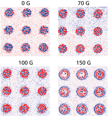
Magnetic force microscopy (MFM) is an important advance in atomic force microscopy that opened up the study of submicrometer magnetic domains. The technique has especially found a central place in the magnetic data storage industry, i.e., in the imaging of magnetic media and devices, whether in the analysis of magnetically recorded bits or in the performance of transducers that read/write them. MFM is also used in fundamental research of magnetic materials and composites, from nanoparticles and nanowires to ferritin proteins. More recently, MFM has been used in tandem with piezoelectric force microscopy (PFM) to characterize multiferroic composites that exhibit magnetoelectric coupling. These composites that consist of both magnetorestrictive and piezoelectric components can also be characterized by operating PFM under an applied in-plane magnetic field using a variable field module (VFM). Research on these novel magnetic materials is driven by the search for higher-density data storage media, high-speed, low-power spintronic devices for computing, and a new class of dual electric-field- and magnetic-field-tunable signal-processing devices.
Ask an AFM expert for more information"Realization of ground state in artificial kagome spin ice via topological defect-driven magnetic writing," J. C. Gartside, D. M. Arroo, D. M. Burn, V. L. Bemmer, A. Moskalenko, L. F. Cohen, and W. R. Branford, Nat. Nanotechnol. 13, 53 (2018). https://doi.org/10.1038/s41565-017-0002-1
"Room temperature uniaxial magnetic anisotropy induced by Fe-islands in the InSe semiconductor van der Waals crystal," F. Moro, M. A. Bhuiyan, Z. R. Kudrynskyi, R. Puttock, O. Kazakova, O. Makarovsky, M. W. Fay, C. Parmenter, Z. D. Kovalyuk, A. J. Fielding, M. Kern, J. van Slageren, and A. Patanè, Adv. Sci. 5, 1800257 (2018). https://doi.org/10.1002/advs.201800257
"Effect of Jahn-Teller distortion on the short range magnetic order in copper ferrite," M. H. Abdellatif, C. Innocenti, I. Liakos, A. Scarpellini, S. Marras, and M. Salerno, J. Magn. Magn. Mater. 424, 402 (2017). https://doi.org/10.1016/j.jmmm.2016.10.110
"Direct visualization of magnetic‐field‐induced magnetoelectric switching in multiferroic aurivillius phase thin films," A. Faraz, T. Maity, M. Schmidt, N. Deepak, S. Roy, M. E. Pemble, R. W. Whatmore, and L. Keeney, J. Am. Ceram. Soc. 100, 975 (2017). https://doi.org/10.1111/jace.14597
"Heat accumulation and all-optical switching by domain wall motion in Co/Pd superlattices," F. Hoveyda, E. Hohenstein, and S. Smadici, J. Phys.: Condens. Matter 29, 225801 (2017). https://doi.org/10.1088/1361-648X/aa6c93
"Ferroelectric control of organic/ferromagnetic spinterface," S. Liang, H. Yang, H. Yang, B. Tao, A. Djeffal, M. Chshiev, W. Huang, X. Li, A. Ferri, R. Desfeux, and S. Mangin, Adv. Mater. 28, 10204 (2016). https://doi.org/10.1002/adma.201603638
"Observation of magnetic anomalies in one-step solvothermally synthesized nickel–cobalt ferrite nanoparticles," G. Datt, M. S. Bishwas, M. M. Rajac, and A. C. Abhyankar, Nanoscale 8, 5200 (2016). https://doi.org/10.1039/c5nr06791j
"G-mode magnetic force microscopy: Separating magnetic and electrostatic interactions using big data analytics," L. Collins, A. Belianinov, R. Proksch, T. Zuo, Y. Zhang, P. K. Liaw, S. V. Kalinin, and S. Jesse, Appl. Phys. Lett. 108, 193103 (2016). https://doi.org/10.1063/1.4948601
"Magnetoelectric quasi-(0-3) nanocomposite heterostructures," Y. Li, Z. Wang, J. Yao, T. Yang, Z. Wang, J.-M. Hu, C. Chen, R. Sun, Z. Tian, J. Li, L.-Q. Chen, and D. Viehland, Nat. Commun. 6, 6680 (2015). https://doi.org/10.1038/ncomms7680
"Patterning magnetic regions in hydrogenated graphene via e‐beam irradiation," W. K. Lee, K. E. Whitener, Jr., J. T. Robinson, and P. E. Sheehan, Adv. Mater. 27, 1774 (2015). https://doi.org/10.1002/adma.201404144
"100-nm-sized magnetic domain reversal by the magneto-electric effect in self-assembled BiFeO3/CoFe2O4 bilayer films," K. Sone, H. Naganuma, M. Ito, T. Miyazaki, T. Nakajima, and S. Okamura, Sci. Rep. 5, 9348 (2015). https://doi.org/10.1038/srep09348
"Design of magnetoelectric coupling in a self-assembled epitaxial nanocomposite via chemical interaction," W. I. Liang, Y. Liu, S. C. Liao, W. C. Wang, H. J. Liu, H. J. Lin, C. T. Chen, C. H. Lai, A. Borisevich, E. Arenholz, J. Li, and Y. H. Chu, J. Mater. Chem. C 2, 811 (2014). https://doi.org/10.1039/c3tc31987c
"Magnetic-field-induced ferroelectric polarization reversal in magnetoelectric composites revealed by piezoresponse force microscopy," H. Miao, X. Zhou, S. Dong, H. Luo, and F. Li, Nanoscale 6, 8515 (2014). https://doi.org/10.1039/c4nr01910e
"Nanocomposite pattern-mediated magnetic interactions for localized deposition of nanomaterials," D. Fragouli, B. Torre, F. Villafiorita-Monteleone, A. Kostopoulou, G. Nanni, A. Falqui, A. Casu, A. Lappas, R. Cingolani, and A. Athanassiou, ACS Appl. Mater. Interfaces 5, 7253 (2013). https://doi.org/10.1021/am401600f
"Micromagnetic modeling of experimental hysteresis loops for heterogeneous electrodeposited cobalt films," M. P. Seymour, I. Wilding, B. Xu, J. I. Mercer, M. L. Plumer, K. M. Poduska, A. Yethiraj, and J. van Lierop, Appl. Phys. Lett. 102, 072403 (2013). https://doi.org/10.1063/1.4793209
"Probing the local strain-mediated magnetoelectric coupling in multiferroic nanocomposites by magnetic field-assisted piezoresponse force microscopy," G. Caruntu, A. Yourdkhani, M. Vopsaroiu, and G. Srinivasan, Nanoscale 4, 3218 (2012). https://doi.org/10.1039/c2nr00064d
"Local characterization of austenite and ferrite phases in duplex stainless steel using MFM and nanoindentation," K. R. Gadelrab, G. Li, M. Chiesa, and T. Souier, J. Mater. Res. 27, 1573 (2012). https://doi.org/10.1557/jmr.2012.99
"Mutual ferromagnetic-ferroelectric coupling in multiferroic copper-doped ZnO," T. S. Herng, M. F. Wong, D. Qi, J. Yi, A. Kumar, A. Huang, F. C. Kartawidjaja, S. Smadici, P. Abbamonte, C. Sánchez-Hanke, S. Shannigrahi, J. M. Xue, J. Wang, Y. P. Feng, A. Rusydi, K. Zeng, and J. Ding, Adv. Mater. 23, 1635 (2011). https://doi.org/10.1002/adma.201004519
"Multiferroic CoFe2O4-Pb(Zr0.52Ti0.48)O3 core-shell nanofibers and their magnetoelectric coupling," S. Xie, F. Ma, Y. Liu, and J. Li, Nanoscale 3, 3152 (2011). https://doi.org/10.1039/c1nr10288e
"Enhanced multiferroic properties and domain structure of La-doped BiFeO3 thin films," F. Yan, T. J. Zhu, M. O. Lai, and L. Lu, Scripta Mater. 63, 780 (2010). https://doi.org/10.1016/j.scriptamat.2010.06.013
"Uniaxial magnetic anisotropy in La0.7Sr0.3MnO3 thin films induced by multiferroic BiFeO3 with striped ferroelectric domains," L. You, C. Lu, P. Yang, G. Han, T. Wu, U. Luders, W. Prellier, K. Yao, L. Chen, and J. Wang, Adv. Mater. 22, 4964 (2010). https://doi.org/10.1002/adma.201001990
"Bimodal magnetic force microscopy: Separation of short and long range forces," J. W. Li, J. P. Cleveland, and R. Proksch, Appl. Phys. Lett. 94, 163118 (2009). https://doi.org/10.1063/1.3126521
"Ion beam sputtered nanostructured semiconductor surfaces as templates for nanomagnet arrays," C. Teichert, J. J. de Miguel, and T. Bobek, J. Phys.: Condens. Matter 21, 224025 (2009). https://doi/org/10.1088/0953-8984/21/22/224025
"Magnetic force microscopy of superparamagnetic nanoparticles," S. Schreiber, M. Savla, D. V. Pelekhov, D. F. Iscru, C. Selcu, P. C. Hammel, and G. Agarwal, Small 4, 270 (2008). https://doi.org/10.1002/smll.200700116
"The band excitation method in scanning probe microscopy for rapid mapping of energy dissipation on the nanoscale," S. Jesse, S. V. Kalinin, R. Proksch, A. P. Baddorf, and B. J. Rodriguez, Nanotechnology 18, 435503 (2007). https://doi.org/10.1088/0957-4484/18/43/435503

© Oxford Instruments 2026