AFM Systems
AFM Accessories
Learning
Contact Us
 Part of the Oxford Instruments Group

Shared instrumentation facilities face unique challenges. Their user base is diverse in both research topics and AFM experience levels. Facility managers must train and support many users and instruments. Limited budgets must be balanced between new equipment purchases and maintaining existing tools. Typical AFMs often worsen these challenges by requiring extensive training, offering limited versatility, and being prone to damage from inexperienced users.
The Jupiter Discovery AFM is different—designed specifically to be much easier to use for new users while offering all the research versatility and ultra-high performance required by more experienced researchers.
Consistent with their mission to train future scientists and engineers, university microscopy core facilities host students who are not only new to AFM but often lack experience with advanced instrumentation. Traditional AFMs pose a steep learning curve, requiring multiple training sessions to achieve basic competence. The Jupiter Discovery flattens this curve with features such as:
Pre-mounted probes make probe exchange quick and easy

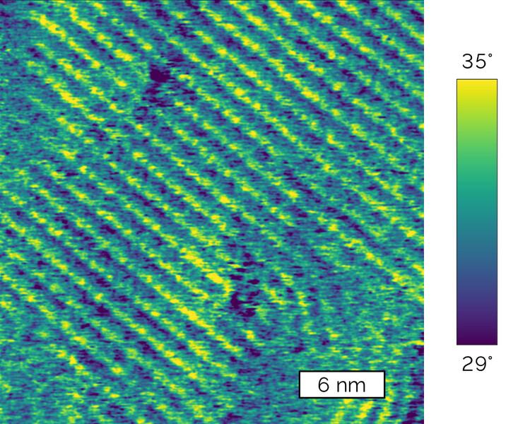
FFM-Topography with AutoPilot readily images the 0.75-nm steps on this silicon carbide substrate (2 µm scan size).
Application Scientist Jason Li shows how easy it is to quickly get high quality images using Jupiter Discovery.
Application Sophia Hohlbauch shows how FFM-Topography with AutoPilot automatically optimizes imaging parameters.
University microscopy core facilities bring together researchers from a wide variety of research fields including 2D materials, semiconductors, polymers, and life sciences. Some users only require basic AFM topography measurements while others need more advanced characterization techniques. Jupiter Discovery covers it all, including:
Jupiter easily resolves the domains, domain boundaries, and even sub-molecular ordering in P3HT, a semiconducting polymer.
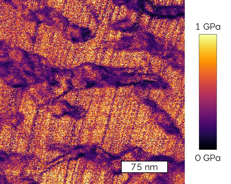
Fast Force Mapping enables mapping the modulus of materials like this PTFE polymer film, distinguishing crystalline regions with higher modulus and amorphous regions with lower modulus.
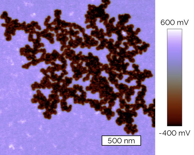
Kelvin Probe Force Microscopy (KPFM) maps the surface potential of samples, here F14H20, a semi-fluorinated alkane molecule that self-assembles into nanostructures.
Core facilities often operate on tight budgets, making it critical to carefully consider the acquisition, operating, and maintenance costs of instruments along with their utilization and usage fees.

Scanner design is highly robust and unlikely to be damaged by mishandling, even by less experienced operators.
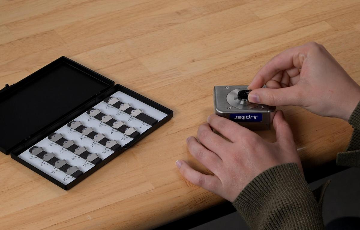
Pre-mounted probes are competitively priced and eliminate the waste that occurs when ordinary probes are accidentally dropped.
If you have any questions, or would like to speak to an expert or request pricing, we will reply to your enquiry shortly.
Get In Touch
© Oxford Instruments 2026