Novel properties of twisted 2D materials are highly dependent on twist angle, with even small differences on the order of fractions of a degree resulting in significant variations in these properties. Twist angle is easily calculated from the periodicity of the twisted 2D sample’s moiré pattern. What is not as trivial is actually producing twisted 2D materials at the desired twist angle, with current fabrication methods suffering from poor accuracy and precision1. As a result, AFM characterisation has become critical in this field. Designed to probe surfaces at the nanoscale and in ambient conditions, high performance AFMs can quickly and easily characterise the moiré of twisted 2D materials.
Torsional Force Microscopy (TFM)2 is one of several AFM imaging modes that can be used to image the moiré of twisted 2D materials. In this mode, the cantilever is maintained at a constant force by the AFM’s height feedback loop while the cantilever is simultaneously excited either mechanically or photothermally at a torsional resonance, as illustrated in Figure 1. A lockin extracts the amplitude and phase of the cantilever’s AC lateral signal, at the torsional resonance. Furthermore, tracking the torsional contact resonance with DART3 can reduce scan line variations and signal drift.
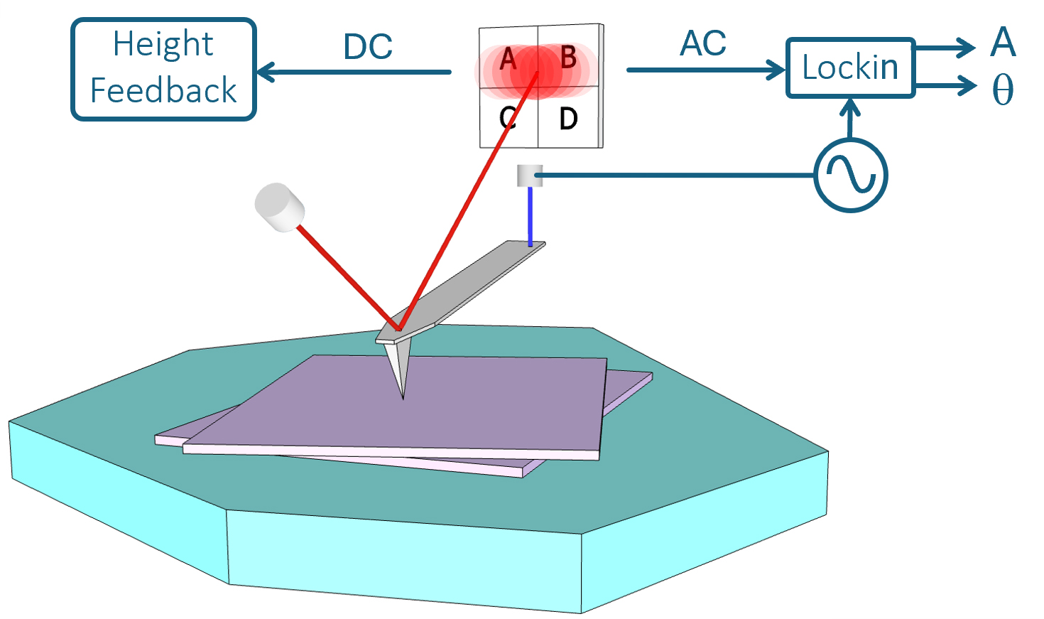
Figure 1: Schematic diagram of TFM. The AFM’s feedback loop maintains the tip�sample force constant while the cantilever is excited at a torsional resonance.
Overview of TFM
TFM is a relatively new technique for imaging the moiré of twisted 2D materials and is thought to be sensitive to dynamic friction at the interface of the AFM tip and sample. As such, it is also able to discern the atomic lattice and thereby identify local crystallographic orientation.
Figure 2 shows a series of successively smaller scans taken in TFM mode on twisted tungsten diselenide (WSe2). The moiré pattern is clearly resolved in each of these scans while the WSe2 lattice is also visible in the two smaller scans. This was imaged with DART TFM using a standard AFM cantilever driven photothermally at its torsional resonance.
Optimal imaging contrast in TFM is often an interplay between the setpoint force, torsional drive amplitude and AFM tip size with blunter AFM tips requiring higher setpoint forces to maintain the contact pressure. Remarkably, keeping the tip sharp is not a requirement for optimal image quality. Indeed, even blunt probes with correspondingly larger imaging setpoint forces of ~1 uN can result in good TFM images, examples of which are shown in this note.
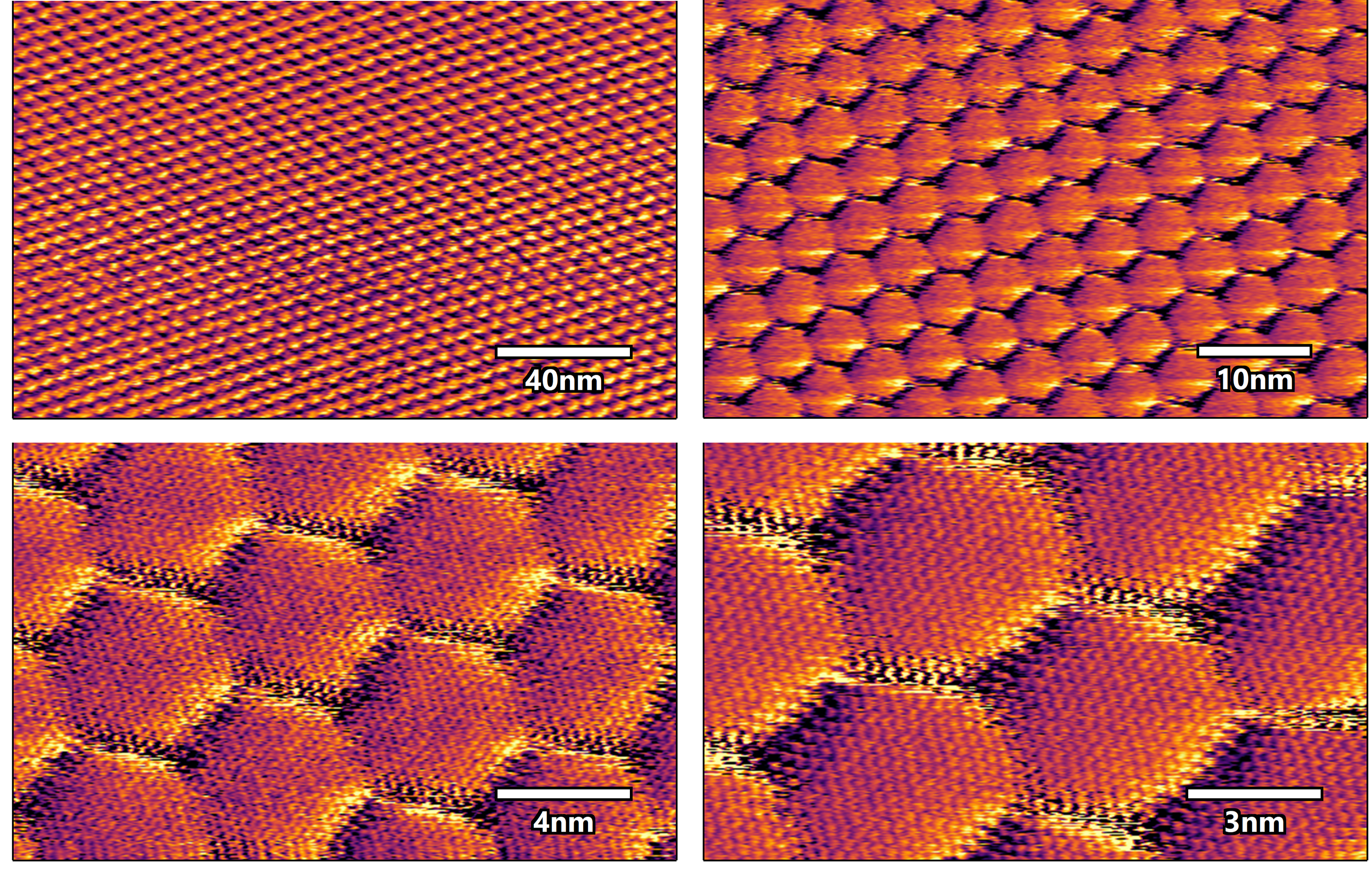
Figure 2: DART TFM Images of twisted WSe2 showing the moiré pattern as well as the atomic lattice in the two smaller scans. Imaged with a Cypher-S and photothermal cantilever excitation. Sample courtesy of the Young lab at UCSB.
Comparison with other imaging modes
Figure 3 shows images of near-magic-angle twisted bilayer graphene (tBLG), in a direct comparison between torsional force microscopy (TFM), piezoresponse force microscopy (PFM) and conductive AFM (cAFM). All three images were taken from the same region of the sample and show that TFM is comparable to these other AFM imaging techniques for imaging tBLG in terms of signal-to-noise and imaging stability.
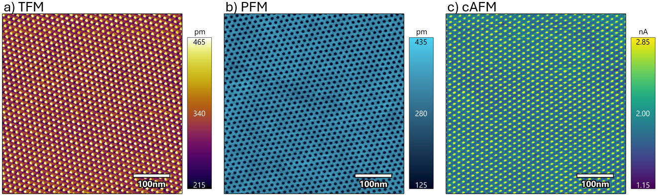
Figure 3: A direct comparison of a) DART torsional force microscopy, b) piezoresponse force microscopy, and c) conductive AFM for imaging the moiré of near-magic-angle twisted bilayer graphene. All scans were taken in the same region of the sample and are comparable in quality in terms of contrast and imaging stability. Imaged with a Cypher-S with an Asyelec.01 cantilever. Sample courtesy of the Young lab at UCSB.
Conclusion
TFM is thought to be sensitive to dynamic friction and can image the moiré of twisted 2D materials as well as the atomic lattice. This technique can give similar contrast and imaging stability compared to PFM and cAFM. Despite being a relatively new mode for imaging the moiré of twisted 2D materials, TFM already shows a lot of potential.
References
1. Lau, Chun Ning, et al. Nature 602.7895 (2022).
2. Pendharkar, Mihir, et al. Proceedings of the National Academy of Sciences 121.10 (2024).
3. Rodriguez, Brian J., et al. Nanotechnology 18.47 (2007).

