Tags: AFM, Atomic Force Microscopy, Scanning Probe Microscopy, SPM, tapping mode
The remarkable properties of nanoscale carbon dots (CDs) make them promising candidates for a range of applications including optical sensing, photovoltaics, and bioimaging. However, successful implementation of these zero-dimensional nanomaterials requires further understanding of their photoluminescence and photo-induced charge transfer processes.
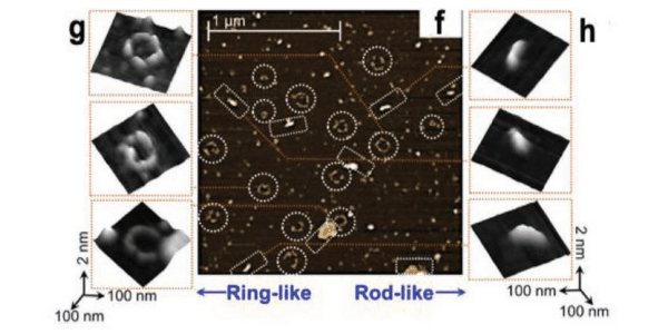 High-resolution AFM images show both rod-like and ring-like structures.
To this end, researchers at the University of Illinois at Urbana-Champaign performed an in-depth investigation of the photophysical properties of CDs on both the single-particle level and in the ensemble-averaged (bulk) state. The CDs were passivated with surface molecules that acted as either electron acceptors or electron donors.
Initial investigation with Transmission Electron Microscopy (TEM) indicated the presence of ring-like structures formed by the association of multiple CDs (see figure below, from the Supplementary Materials). However, the particle diameters measured with TEM deviated from those determined by dynamic light scattering, perhaps due to artifacts induced by TEM sample preparation. The researchers then turned to their Cypher Atomic Force Microscope (AFM) to achieve higher resolution and quantitative particle heights. The high-resolution AFM images revealed both ring-like and rod-like structures. The ring-like structures observed by AFM were more uniform in size and shape compared to those observed with TEM.
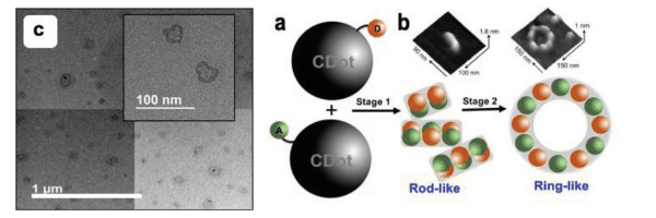
(Left) TEM image of ring-like structures, (Right) AFM images and proposed schematic of CD assembly
Additional experiments were performed with a variety of techniques and combined with density functional theory (DFT) calculations. Other measurements showed the type of structure formed depended on experimental variables including time, particle concentration, and hydrogen bonding. From their observations, the researchers concluded that the bulk-state photophysical properties could be affected by formation of hierarchical structural assemblies of CDs.
The findings provide insight into the mechanisms of photo-induced emission in CDs. As a result, they may accelerate the use of these nanomaterials in next-generation optoelectronic and quantum technology.
Instrument used
Cypher Atomic Force Microscope (AFM)
This research article highlights the high-resolution imaging of a Cypher AFM. The Cypher family of AFM’s have been designed to achieve higher spatial resolution than most AFM’s. This performance not only improves image quality, but also makes it far easier to obtain high-quality data consistently. The result is greater productivity in generating high-resolution, publication-quality data in your lab. This impressive performance is engineered into the Cypher from the ground up and informed by many decades of collective AFM experience among Asylum scientists and engineers.
-20210210011323.png)
It begins with an ultra-stable mechanical design. The most critical mechanical design feature of an AFM is the rigidity of the structure that connects between the sample and the probe. Any relative motion in this structure except for the intended scan waveform compromises imaging quality. In Cypher AFMs, this “mechanical loop” is contained entirely within the scanner, which enables it to be very short and very rigid. This is what enables the incredible height noise of <15 pm on Cypher—which is less than half that of other common AFMs.
The performance of the scanner in both the XY axes and Z axis is also a critical factor in imaging performance. This is influenced by both the mechanical and electronic design of the scanner and its integrated position sensors. Scanners in our Cypher AFM’s use a monolithic direct-drive flexure design that increases its stiffness (i.e., resonance frequency), which makes them lower noise, faster, and more immune to external noise sources (e.g. building vibrations). Asylum is also the only AFM company that uses linear variable differential transformers (LVDTs) as position sensors in our scanners. Compared to more commonly used capacitive sensors and strain gauge sensors, LVDTs offer lower noise, lower drift, inherently linear response, and never need to be recalibrated. Cypher’s industry leading 60 pm XY sensor noise and 50 pm Z sensor noise enable ultra-high resolution imaging along with highly accurate metrology.
The AFM probe and deflection sensing optics are the next critical component since it is here that topography and properties are measured as the tip scans the sample. Cypher AFMs are compatible with the very smallest commercial AFM cantilevers, which enable not only faster scanning but also higher resolution imaging. Compatibility with these cantilevers requires a small laser spot focus from the deflection sensing optics. Cypher is the only commercial AFM with user-interchangeable laser modules for optimal performance on small cantilevers and conventional cantilevers and for both imaging and force-distance curves and force mapping.
Finally, the system electronics are the last determining factor in AFM imaging performance. Even very small electronic noise can result in higher noise in AFM images. So every component in both the analog and digital design of the control electronics is designed and scrutinized to ensure that they don’t compromise imaging performance.
Overall AFM performance is determined by a complex combination of these factors. These have all been optimized in Cypher family of AFM’s, which cannot be summarized by a single specification in a datasheet. That’s why we encourage you to evaluate Cypher performance in terms of actual results, whether those are in a sales demonstration, your lab, or in published results like the ones shared here.
Techniques used
Samples were made by drop-casting mixtures of donor and acceptor CDs in water onto freshly cleaved mica. After drying overnight, the samples were imaged in ambient conditions on a Cypher AFM in tapping mode. Due to the Cypher’s exceptional spatial resolution, the resulting topography images enabled precise identification and characterization of individual ring-like and rod structures.
Citation: I. Srivastava, J. Khamo, S. Pandit et al., Influence of electron acceptor and electron donor on the photophysical properties of carbon dots: A comparative investigation at the bulk‐state and single‐particle level. Adv. Funct. Mater. 29, 1902466 (2019). https://doi.org/10.1002/adfm.201902466
Note: The data shown here are reused under fair use from the original article, which can be accessed through the article link above.

