AFM Systems
AFM Accessories
Learning
Contact Us
 Part of the Oxford Instruments Group
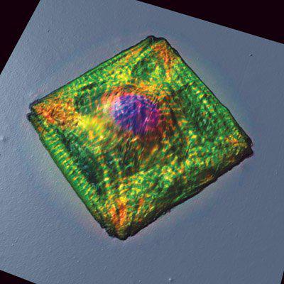
A rat cardiac myocyte was cultured on a micro-contact-printed square island of extra-cellular matrix (ECM) protein on a PDMS substrate. The cell was fixed and stained for actin (green), sarcomeric alpha-actinin (red) and DNA (blue). After fixation, the cell was scanned by AFM. Merged topography and fluorescent images reveal that the actin/alpha-actinin myofibrils and the nucleus are responsible for specific AFM topography. The 92 µm image was taken by the Disease Biophysics Group at the School of Engineering and Applied Sciences, Harvard University, headed by Prof. Kevin Kit Parker. Image courtesy of K. Parker and N. Geisse, Harvard University. Control of Myocyte Remodeling In Vitro with Engineered Substrates Geisse NA, Sheehy SP, Parker KK In Vitro Cell Dev Biol Anim. 2009. 45(7):343-50.

© Oxford Instruments 2026