AFM Systems
AFM Accessories
Learning
Contact Us
 Part of the Oxford Instruments Group
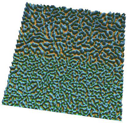
The image is a surface plot of a 1 micron square of nanoparticle network. The top half of the image has been scanned repeatedly with the AFM over an eight hour period, while the bottom half of the image was scanned just once in order to obtain this image. This technique can be used to gain direct control over the length scales of nanoparticle structures. Imaged with the MFP-3D AFM. Image courtesy of M. Blunt and P. Moriarty, University of Nottingham.

© Oxford Instruments 2026