AFM Systems
AFM Accessories
Learning
Contact Us
 Part of the Oxford Instruments Group
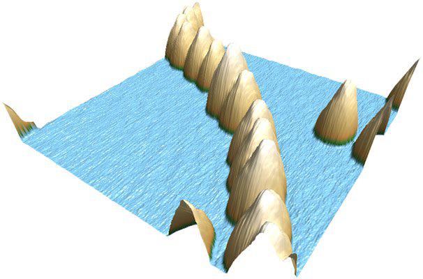
3D rendering of an AFM topographical image, 450 nm scan of 12 nm Au nanoparticles (NP) on a wafer-grade polished glass substrate. The NPs were initially aligned using insulin fibers as an adherent sacrificial biotemplate. Fibers were subsequently removed by exposure to a low-pressure plasma, leaving the Au NPs in their template positions. Imaged with the MFP-3D AFM. Image courtesy of Shuchen Hsieh and Chiung-Wen Hsieh, National Sun Yat-sen University, Taiwan ROC.

© Oxford Instruments 2026