AFM Systems
AFM Accessories
Learning
Contact Us
 Part of the Oxford Instruments Group

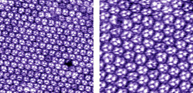
Two closed loop images of the cytoplasmic face of bacteriorhodopsin in buffer, 110 nm (left) and 50 nm (right) scans. The left scan shows a missing protein from the lattice. Both images were taken with the Cypher AFM. Images courtesy of G.M. King Lab, Univ. of Missouri-Columbia.
Date: 16th November 17
Last Updated: February 20, 2019, 5:23 pm
Author: Asylum Research
Category: Asylum Gallery Image
