AFM Systems
AFM Accessories
Learning
Contact Us
 Part of the Oxford Instruments Group
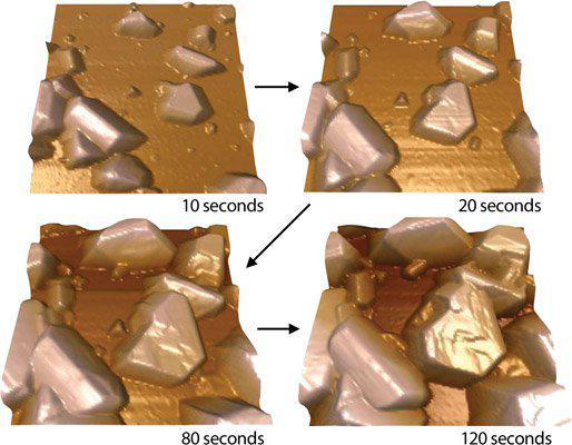
AFM images of the Boron Doped Diamond electrode surface (left) before deposition and (right) after a 120 s deposition of Bismuth nanoparticles at -1.2 V. 2.5 µm scan. Imaged with the MFP-3D AFM. Image courtesy of R. Compton, Oxford University.

© Oxford Instruments 2026