AFM Systems
AFM Accessories
Learning
Contact Us
 Part of the Oxford Instruments Group

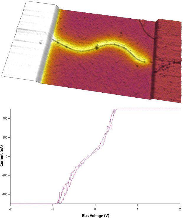
Electric force microscopy (EFM) (top) and a current-voltage curve (bottom) of a carbon nanotube attached to an electrode. Since the carbon nanotube is conducting, and has continuity to ground here, the EFM phase image shows excellent contrast. In the image, color represents the EFM phase channel, and the surface rendered with ARgyle represents topography. Scan size 5 µm x 2.5 µm. Imaged with the MFP-3D AFM. Courtesy Minot Lab, Oregon State University.
Date: 16th November 17
Last Updated: July 12, 2018, 11:13 am
Author: Asylum Research
Category: Asylum Gallery Image
