AFM Systems
AFM Accessories
Learning
Contact Us
 Part of the Oxford Instruments Group
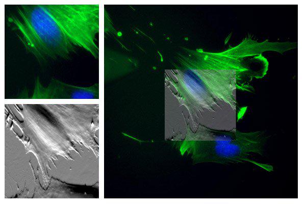
Upper Left: Fluorescent image of cells labeled with DAPI (nucleus) and Alexa Fluor 488 (actin filaments) and viewed with wide-fluorescence using the Nikon TE2000U inverted optical microscope and a 40x objective. Lower Left: AFM deflection image, 60 µm scan. Right: AFM deflection data (50% transparency) overlaid onto merged fluorescence optical image.

© Oxford Instruments 2026