AFM Systems
AFM Accessories
Learning
Contact Us
 Part of the Oxford Instruments Group
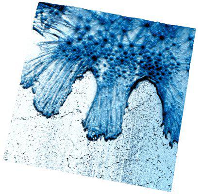
Dynamic AFM image of a live rat mesenchymal stem cell showing both linear and geodesic arrangements of the F-actin cytoskeleton. The high energy dissipation indicated at the geodesic vertices confirms the prediction of Lazarides (J. Cell. Biol. 68, 202-219 (1976)) that the vertices should have higher flexibility based on the absence of tropomyosin and myosin at these junctions. 90 µm scan. For more information, see HFSP Journal, 1, 181-191 (2007). Image courtesy of Suzi Jarvis and colleagues, Conway Institute for Biomolecular and Biomedical Research, University College Dublin.

© Oxford Instruments 2026