AFM Systems
AFM Accessories
Learning
Contact Us
 Part of the Oxford Instruments Group
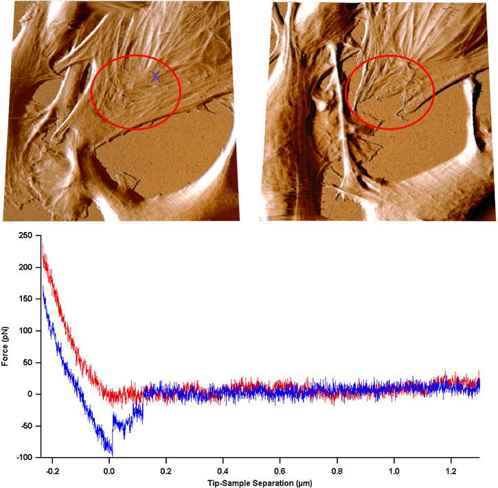
Deflection images of living outer Annulus cells from Bovine Intervertebral discs, day 6 (after seeding), imaged in culture medium containing 10% fetal bovine serum (FBS) using Contact Mode. Left image was acquired before a series of force curves were taken on the area of the cell that is circled in red. Right image was acquired 45 minutes later. Displayed force curve was acquired on the cell marked with an ?X?. It can be clearly seen that the cells have moved, undergone cytoskeletal reorganization and, where the force curves were taken, were damaged by the AFM tip, 54 µm. Data courtesy of Y. Dror and J. Klein, Oxford University and Weizmann Institute, NanoInteract consortium.

© Oxford Instruments 2026