AFM Systems
AFM Accessories
Learning
Contact Us
 Part of the Oxford Instruments Group
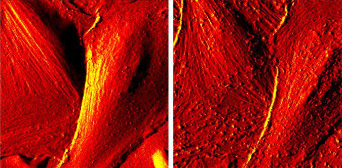
Before (left) and after (right) chemical treatment with mannitol. Living GP8 rat cerebral endothelial cells cultured until confluency in a petri dish coated with rat tail collagen in DMEM/F12 (Dulbecco's Modified Eagles Medium with F12 salt), supplemented with 12% PDS (Plasma Derived Serum). They were imaged at a temperature around 31°C. The left image is before chemical treatment. After imaging, without moving the sample and the AFM head, the medium was exchanged with the same solution, containing 0.55 M mannitol which has a reversible hyperosmotic effect at this concentration. After changing the solution, the same cells were imaged to see the mannitol induced changes (right image). 40 µm scans. Image courtesy of Z. Bálint and G. Váró, Hungarian Academy of Sciences.

© Oxford Instruments 2026