AFM Systems
AFM Accessories
Learning
Contact Us
 Part of the Oxford Instruments Group
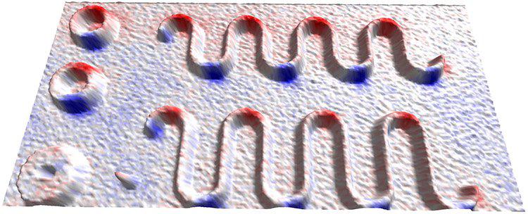
Magnetic Force Microscopy (MFM) phase data overlaid on topography of cobalt nanoribbons composed of half-circles with 800 nm radius connected by straight sections of length 800 nm (top) and 1600 nm (bottom). The sample is in the "onion" state with head-to-head (blue) and tail-to-tail (red) domain walls that have been created by the application and removal of a uniaxial in-plane magnetic field using the Variable Field Module (VFM) on a MFP-3D AFM. Scan size 10 µm x 20 µm. Image courtesy of Jessica Bickel and Kathy Aidala, Mount Holyoke College.

© Oxford Instruments 2026