AFM Systems
AFM Accessories
Learning
Contact Us
 Part of the Oxford Instruments Group

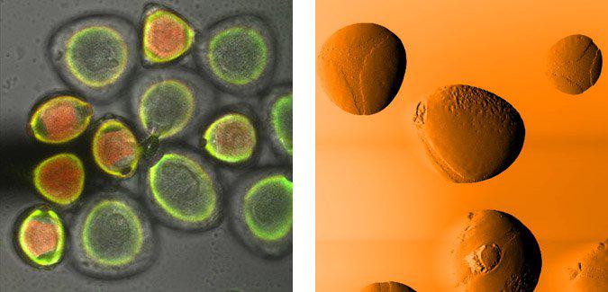
Left: Pollens were imaged with the Olympus FluoView 1000. Confocal fluorescence (red, green) with transmitted light (grayscale). Shadow of the AFM cantilever is at the middle left of the image, 50 µm scan. Right: Pollens were imaged with the MFP-3D-CF. Amplitude channel. Due to the height of the pollen grains, only the top surfaces of the tallest grains are in range, 96 µm scan.
Date: 16th November 17
Last Updated: February 20, 2019, 5:23 pm
Author: Asylum Research
Category: Asylum Gallery Image
