AFM Systems
AFM Accessories
Learning
Contact Us
 Part of the Oxford Instruments Group

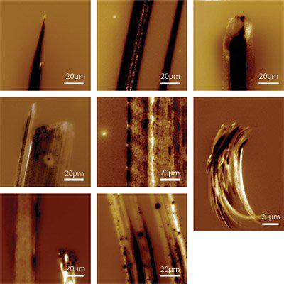
Gallery of various surface potential AFM images illustrating the charge patterns on polymer films generated by rubbing polymer covered cotton swabs across the surface (top row: PAAm rubbed by COC). The rubbing direction is from top to bottom. The lower right shows a complete trace of PVAc rubbed by PAAm (110 µm x 162 µm composed of two separate scans). The color scale varies slightly from image to image but in all images, the potential signal covers a range of a few volts. Imaged with the MFP-3D AFM, 90 µm scan size.
Date: 16th November 17
Last Updated: July 12, 2018, 11:13 am
Author: Asylum Research
Category: Asylum Gallery Image
