AFM Systems
AFM Accessories
Learning
Contact Us
 Part of the Oxford Instruments Group

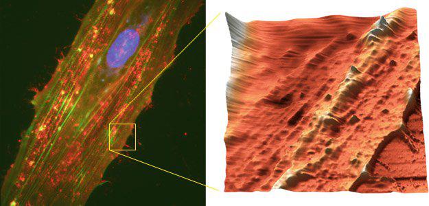
Left: Fixed endothelial cells (HUVECC cell line) were labeled with Hoescht 33342 (nucleus), Alexa Fluor 488 (actin filaments) and Alexa Fluor 546 (ICAM-1) and viewed with wide-field fluorescence using the Nikon TE2000U inverted microscope and a 40x objective. Right: The AFM image was scanned at the area indicated by the box and then rendered in 3D, 35 µm scan. Alexa Fluor 546 and antibody courtesy of R. Waugh and E. Lomakina, University of Rochester.
Date: 16th November 17
Last Updated: July 12, 2018, 11:13 am
Author: Asylum Research
Category: Asylum Gallery Image
