AFM Systems
AFM Accessories
Learning
Contact Us
 Part of the Oxford Instruments Group
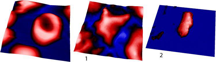
Red blood cell on a mica substrate imaged in PBS solution (left). The cell displays the characteristic biconcave shape of healthy RBCs. It has a diameter of approximately 8µm and a height of approximately 2.5 µm, 20 µm scan. Sample purchased from Research Blood Components, Brighton, MA. Red blood cells from a patient with sickle cell disease (SCD) imaged on a mica substrate in PBS solution. RBC (1), 25 µm scan, is in an oxygenated environment while RBC (2), 20 µm scan, is in the fully deoxygenated state. These RBCs have a highly irregular morphology as compared to the characteristic biconcave shape of normal RBCs. Polymerized hemoglobin within the cell results in highly irregular morphologies characterized by protrusions, elongation, and bumps. Sample courtesy of Biree Andemariam, MD, University of Connecticut Health Center. All images courtesy of Jamie Maciaszek and Professor George Lykotrafitis, University of Connecticut, and adapted from: Maciaszek, J L, B Andemariam, and G Lykotrafitis. "Microelasticity of Red Blood Cells in Sickle Cell Disease." The Journal of Strain Analysis for Engineering Design 46, 5 (2011): doi:10.1177/0309324711398809. Maciaszek, Jamie L, and George Lykotrafitis. "Sickle Cell Trait Human Erythrocytes Are Significantly Stiffer Than Normal." Journal of Biomechanics 44, 4 (2011): doi:10.1016/j.jbiomech.2010.11.008.

© Oxford Instruments 2026