Tags: Scanning Probe Microscopy, high resolution, High Resolution, atomic resolution, Atomic Force Microscopy, AFM
January is the “resolution” time of year, and one I’ve been intending to make for a long time is to take time out from my schedule and do something I truly enjoy – learn what our creative and talented customers have been up to. One of the delightful rewards of manufacturing scientific instruments is seeing all the exciting ways our excellent customers use them.
At the last Multifrequency AFM Conference in Madrid [i], I was lucky enough to attend Kristen Burson’s talk, “Resolving 2D Amorphous Materials with Atomic Force Microscopy.” She had been using a Cypher AFM at the Fritz Haber Institute in Berlin for high-resolution imaging of an amorphous silica bilayer. Her and her coworkers’ results have also appeared in Applied Physics Letters [ii].
I thought this work was really nice for a lot of reasons:
1. Many years ago as a postdoc I tried to image glass with no success, so I was excited to see the amorphous surface imaged in liquid with our Cypher ES system.
2. Glass is a fascinating material with a range of mechanical and electrical properties.
3. It seemed like an ideal material for testing the resolution of an AFM since, unlike a calcite or other crystal, it is essentially nothing but a collection of defects.
4. Glass is ubiquitous. I happened to use a common glass slide for the images below, but it will be fun to try out other glasses.
I didn’t have access to Kristen’s bilayers, but there is a lot of glass sitting around the lab. I started simple, with a microscope slide manufactured from common soda-lime glass, distributed by Fischer Scientific. Unlike calcite, glass is not self-cleaning, so some care needs to be taken with the sample prep and handling. However, in the five attempts I took at achieving amorphous lattice resolution, I was successful 100% of the time with a simple protocol:
1. Vigorously scrub the glass surface with a laboratory dishwashing detergent. Don’t be afraid of using elbow or “thumb” grease – really give it a scrubbing.
2. Rinse copiously with distilled water.
3. Blow dry with dry nitrogen.
4. Put a droplet of the imaging solution (in this case, ultrapure distilled water) onto the glass. It should spread out evenly, demonstrating that the glass surface is clean and “hydrophilic.”
For imaging conditions, I chose to use a Nanoworld Arrow UHF cantilever in tapping mode using a free amplitude of ~4 nm and a setpoint of ~2 nm. As in Burson et al.’s work, I used blueDrive photothermal excitation for a stable, well-controlled amplitude. Typical scan ranges were 5-10 nm. The glass slides were not particularly smooth, but it was generally easy to find smooth hilltops or valleys where the amorphous structure could be resolved. A few examples are shown below.
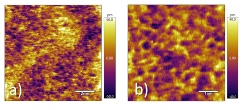
Figure 1. Topography images of disordered lattice imaged at an amplitude setpoint of 2 nm. a) 10 nm scan and b) 5 nm scan. Both images clearly demonstrate sub-nm amorphous glass surface.
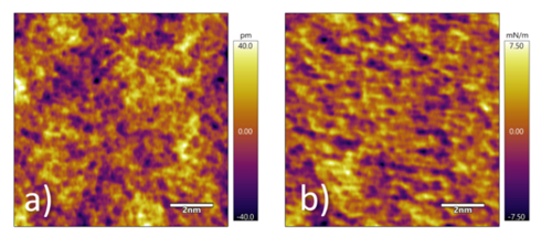
Figure 2. a) Surface topography and b) tip-sample stiffness of a region of the glass sample imaged using AMFM stiffness mapping. 10 nm scan
Using blueDrive and the Arrow UHF, it was also possible to simultaneously map the topography and tip-sample stiffness using AM-FM mode (Figure 2). Like Burson et al., we saw a disordered-appearing surface, with length scales similar to those reported in that paper. Interestingly, these structures were visible with slightly different resolutions with every attempt I made. This is a testament to the low noise of the Cypher AFM and to the reliable sharpness of the Arrow UHF cantilevers. For more information on noise, my colleague Jason Cleveland put on a really nice webinar a few years back that is still available. Click here for a link to the webinar.
Thanks for reading. 2018 has just begun; I’m hopeful I’ll be able to keep this New Year’s resolution of reviewing other cool customer work.
Happy New Year and all the best,
Roger

References
[i] The next Multifrequency AFM meeting is this April in Madrid – see http://www.icmm.csic.es/multifrequency-afm/ for more info.
[ii] Kristen M. Burson, Leonard Gura, Burkhard Kell, Christin Büchner, Adrian L. Lewandowski, Markus Heyde, and Hans-Joachim Freund, Applied Physics Letters 108, 201602 (2016); http://aip.scitation.org/doi/abs/10.1063/1.4949556.

