Tags: AFM, Atomic Force Microscopy, nanolithography, Scanning Probe Microscopy, SPM, tapping mode
A research team at the University of Illinois created a graphene-based sensor to detect oxygen permeation across lung membranes. AFM images of samples in healthy and diseased states helped them uncover relations between oxygen transport and membrane structure.
Pulmonary membranes are thin layers of lipid-protein complexes at the air/liquid interface of respiratory air sacs (alveoli) that mediate the exchange of oxygen and carbon dioxide in the lungs. However a detailed understanding of this mechanism, especially how it is affected by diseases such as bacterial pneumonia, remains elusive.
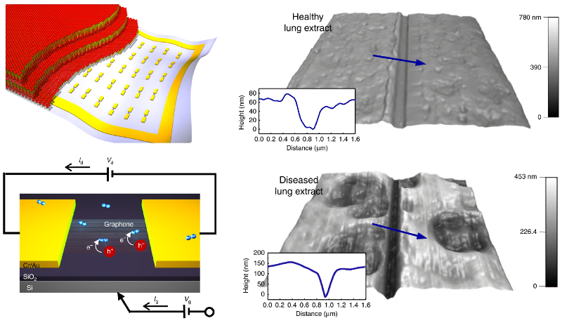
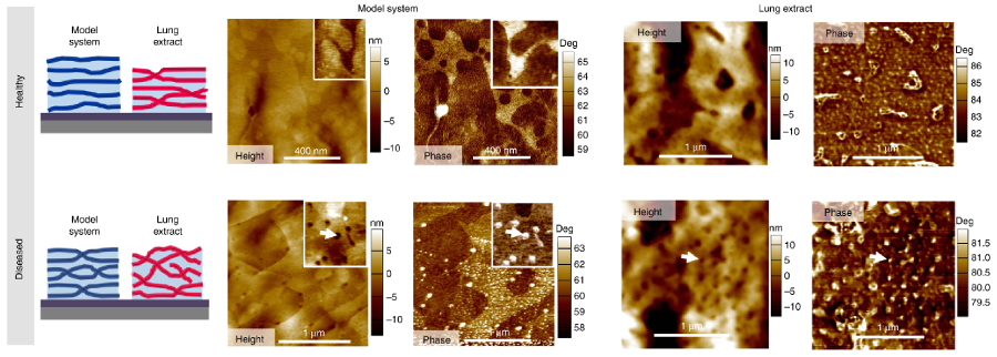
To investigate these issues, researchers developed a micrometer-scale sensor based on graphene field-effect transistors (FETs). By directly coating the devices with thin lung membranes, they measured oxygen permeation through the membranes. Comparison of the results with x-ray and AFM data revealed a direct connection between oxygen transport and structural transformations in diseased and diseased-mimetic samples. In particular, AFM images of nanoscale morphology and compositional contrast identified greater numbers of membrane contacts, or stalks, in diseased states that promoted an increase in oxygen permeation.
The results of this work bring insight into lung membrane function and thus could advance our understanding and treatment of a range of respiratory diseases.
Instrument used
MFP-3D AFM
Techniques used
Tapping mode images of topography and phase were acquired in humid environments (up to 98% RH). The high spatial resolution of MFP-3D AFMs enabled accurate topographical measurements for evaluating specimen morphology, while phase image contrast gave information about variations in mechanical properties. The experiments also used the MFP-3D’s nanolithography capabilities to scratch through the thin samples so that their thickness could be determined.
Citation: M. Kim, M. Porras-Gomez, and C. Leal, Graphene-based sensing of oxygen transport through pulmonary membranes. Nat. Commun. 11, 1103 (2020). https://doi.org/10.1038/s41467-020-14825-9
Note: The original article featured above was published as Open Access. The data shown here are reused under fair use and under the Creative Commons license of the original article, which can be accessed through the article link above.

