Tags: AFM, Atomic Force Microscopy, Scanning Probe Microscopy, SPM, tapping mode
Japanese researchers investigated structural transformations on surfaces of self-assembled coordination polymers with AFM. Liquid-phase imaging with crystal lattice resolution revealed sharp, reversible changes to the polymer framework in the presence of guest molecules.
Porous coordination polymers (PCPs) are self-assembled frameworks of organic ligands and metal ions. Some PCPs can reversibly alter their structure, yet remain in crystalline form, when exposed to guest molecules or external stimuli. This remarkable ability offers exciting possibilities for sensing, separation, and storage of molecules.
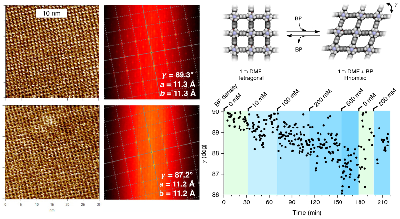
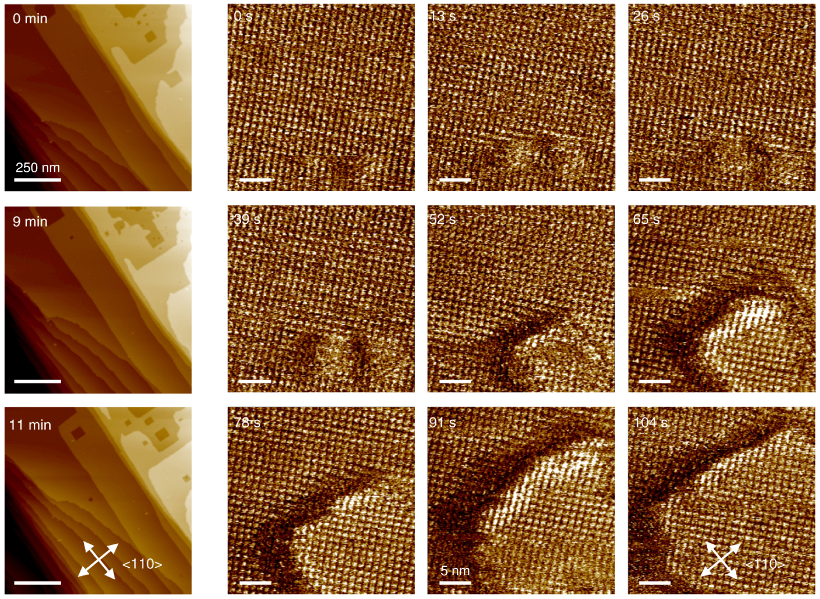
Researchers at Kyoto University used AFM to gain insight into these processes at surfaces of the PCP (Zn2(1,4-ndc)2(dabco))n, where 1,4-ndc is 1,4-naphthalenedicarboxylic acid and dabco is 1,4-diazabicyclo[2.2.2]octane. Lattice-resolution images acquired in dimethylformamide (DMF) solution showed the lattice suddenly changed from tetragonal to rhombic symmetry in the presence of biphenyl (BP) molecules. The switch occurred even at guest concentrations much lower than would trigger the same transformation in bulk crystals. In addition, layer-by layer delamination events were visualized in real time by acquiring a new image every few seconds.
The results provide new details about the highly responsive nature of PCP surfaces and their structural transformations that could accelerate development of new molecular devices.
Instrument used
Cypher ES with Liquid Perfusion Cell
Techniques used
Images were acquired in tapping mode on the Cypher ES AFM. With use of its environmental control capabilities and the Liquid Perfusion Cell, experiments were performed in solution at a constant temperature of 28 °C. Even in liquid, stable images with lattice-scale resolution were obtained due to the superb resolution of Cypher AFMs and its blueDrive tapping mode technology. The high fidelity of the resulting images meant that 2D autocorrelation analysis yielded precise values of the rhombic angle γ. In addition, the fast scan rates of Cypher AFMs made it possible to capture an image every ~13 seconds so as to visualize transformation dynamics.
Citation: N. Hosono, A. Terashima, S. Kusaka et al., Highly responsive nature of porous coordination polymer surfaces imaged by in situ atomic force microscopy. Nat. Chem. 11, 109 (2019). https://doi.org/10.1038/s41557-018-0170-0
Note: The data shown here are reused under fair use from the original article, which can be accessed through the article link above.

