Teijin Ltd is a Japanese company that provides innovative solutions in four business areas, including its two core businesses in Materials and Healthcare, as well as Fibers & Related Products, and IT. Their corporate philosophy is “enhancing the quality of life.” To maintain its leadership in materials development, Teijin recently acquired Oxford Instruments’ Jupiter XR Atomic Force Microscope (AFM) at its Iwakuni Development Center. In this interview, we asked Dr. Bunsow Nagasaka and Mr. Hirofumi Kurimoto at the Teijin Material Analysis Research Center to discuss the Jupiter XR and their reasons for choosing the Oxford Instruments solution from among 19 other AFM companies.
Teijin uses the Jupiter XR to analyze and evaluate a wide range of samples for the development of materials in pharmaceuticals, medical materials, polymers, and inorganics (specifically carbon fibers).
Teijin’s Tokyo Research Center in Hino city acquired the company’s first AFM over 20 years ago. At that time, AFM was a more of a novelty, and was quite interesting because they could obtain brown-colored surface profile images and phase images by AFM measurement. However, samples had to be extremely flat, the AFM was susceptible to vibration, and measurements were very time-consuming, so trying to measure actual samples was a nearly impossible task. In addition, the electron microscope was the optimum method at that time for acquiring surface morphology images. Since we were able to interpret the crystalline and amorphous portions of polymer materials by the shape plus the contrast of backscattered and transmitted electron images, we realized that running the AFM back then was not as useful as we had hoped. However, we still needed to evaluate surface irregularities of individual fibers. Measuring the roughness of a surface with curvature is extremely difficult, even with a confocal laser microscope, so AFM would have been the best technique for characterizing the fiber structure.
Fast forward 20 years, and the need to get more detailed structural information of the fibers became increasingly important. As we began investigating the best way to upgrade our old AFM, we learned that the Z range of the AFM, that is, the operating range in the height direction, is much larger in modern AFMs than in the past. I thought finally it might be possible to measure and evaluate the surface of our fibers! For the demonstration, we prepared cross-sectional samples by cryogenic ion milling from a polymer alloy cut and sent them to several AFM manufacturers. This was a cross section of a polymer alloy with phase separation of the polyamide and epoxy phases and the key point here was whether the instrument could correctly characterize the structure (shape image) and the elastic modulus, (which shows the hardness distribution). When we received the first demo report from another AFM manufacturer, there was noise in the surface profile and the hard and soft parts of the sample were reversed in the elastic modulus image, giving us the impression that AFMs hadn’t changed much after all. That AFM company commented that the roughness of the sample surface was much too large, so the sample itself was not good. I was discouraged at the time, thinking that, after all, this kind of measurement would be difficult with AFM.
However, when we received the elastic modulus map and data acquired with the Jupiter AFM, the hard polymer and soft polymer regions were displayed correctly and the modulus values were accurate. Frankly, I had almost given up hope that AFM could be useful for us, but the data Oxford sent us was on a completely different level. And that is when I changed my opinion that AFM technology has evolved. To tell the truth, the person in charge of preparing the cross sections didn’t have enough time to do the final touches on the sample, so I thought it was amazing that Oxford could acquire data even under these challenging sample conditions. I was astonished by the high quality of the data that Oxford submitted. The noise in the measurements was at a completely different level and Oxford’s blueDrive photothermal cantilever excitation technology was very useful.
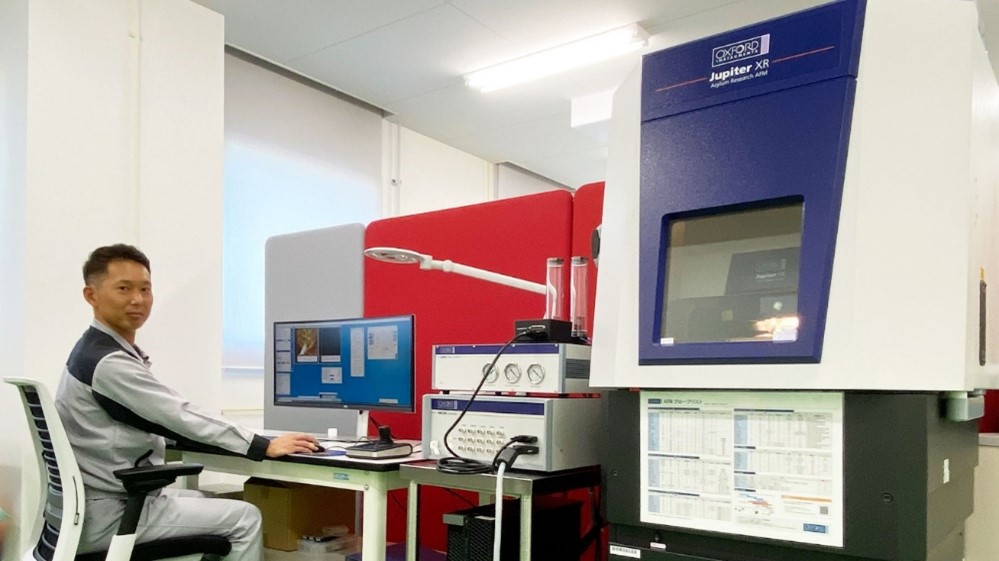
One of the most important points for us regarding the AFM was to know whether we would be able to perform the measurements again by ourselves. Mr. Kurimoto, who is basically a beginner with AFM, was able to perform the measurements and obtain correct results. In comparison with conventional AFM used in the past, the Jupiter XR can acquire images in a short time with good repeatability. For example, when measuring the surface of our fibers in conventional AFM, we were barely able to see the shape, but with the Jupiter XR, it is possible to observe the detailed surface morphology. Because cantilever tuning is also very easy on the Jupiter, the AFM can complete an analysis in half the time required, or at about twice the speed. The Jupiter XR is such a high performance instrument that it is in almost full operation every day.
The Oxford AFM application team is really excellent. They are outstanding. Unfortunately, our company doesn’t have experts who have used AFM since graduate school so I think that whether we can receive technical support from the maker’s staff was also a critical factor in our selection process. For example, probe selection is crucial for AFM measurements, but there are so many types available that it is impossible to know which to choose. With Oxford, we receive accurate and useful advice from expert staff about which probes would be ideal for our applications. This may be an exaggeration, but without Oxford’s application staff, we might not have purchased this instrument. I am extremely satisfied with the performance of the Jupiter AFM and the technical knowledge of Oxford’s AFM team, and I have turned into a total fan of Oxford Instruments.
-20230605131155.png)

