AFM Systems
AFM Accessories
Learning
Contact Us
 Part of the Oxford Instruments Group
British researchers studied catalytic processes using in-situ AFM imaging with single-molecule resolution. They observed spontaneous formation of methanol–water structures on the surface of HOPG, with the catalysis rate enhanced by electric fields.
Methanol is an appealing option for cleaner fuel storage and transportation that is created by catalysis of volatile organics and water. However, more information about the molecular-scale details of catalytic processes is needed to improve current production methods and to develop simpler, cheaper ones.
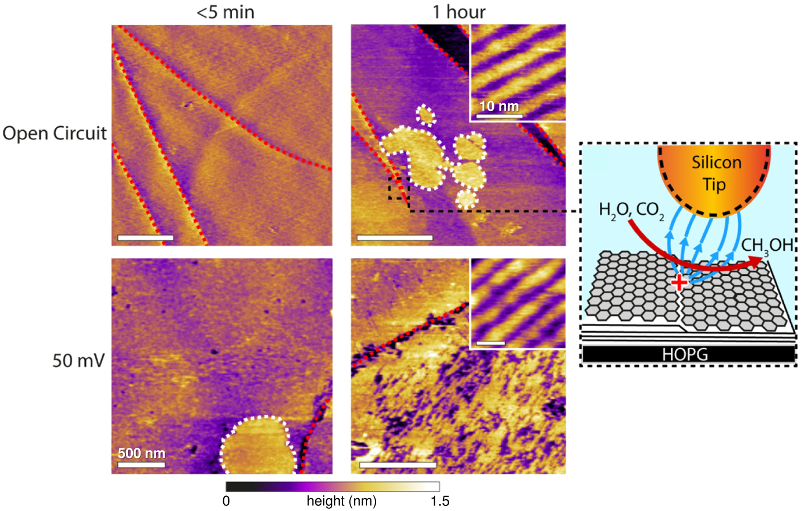
Researchers at Durham University tackled this topic using lattice-scale AFM imaging and nuclear magnetic resonance (NMR). They found that highly oriented pyrolytic graphite (HOPG) spontaneously catalyzed methanol at room temperature when immersed in ultrapure water.
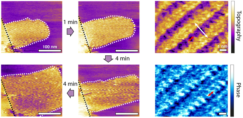
The images revealed methanol–water interfacial structures (white dashed areas in figures) containing row-like features with nanometer-scale periodicity. These nanostructures formed at catalytically active surface features such as atomic step edges (red dashes). Experiments also indicated that catalysis rates increased significantly in the presence of electric fields, even those induced by the tiny AFM tip.
The results provide valuable insight that could aid in developing new organic catalysts and novel carbon-based nanodevices.
In-situ AFM experiments were performed in tapping mode on a Cypher ES AFM. The environmental control capabilities of the Cypher ES allowed high-resolution images to be acquired in water at constant temperature (40 °C). Stable images of molecularly-ordered structures were made possible by Cypher’s exceptional spatial resolution, due in part to its blueDrive tapping mode technology. In fact, the high image quality enabled precise measurements of periodicity: 0.79 ± 0.08 nm parallel and 2.45 ± 0.08 nm perpendicular to the main rows (red and white arrows, respectively, in top figure).
Citation: W. Foster, J. Aguilar, H. Kusumaatmaja, and K. Voϊtchovsky, In situ molecular-level observation of methanol catalysis at the water–graphite interface. ACS Appl. Mater. Interfaces 10, 34265 (2018). https://doi.org/10.1021/acsami.8b12113
Note: The data shown here are reused under fair use from the original article, which can be accessed through the article link above.

© Oxford Instruments 2026